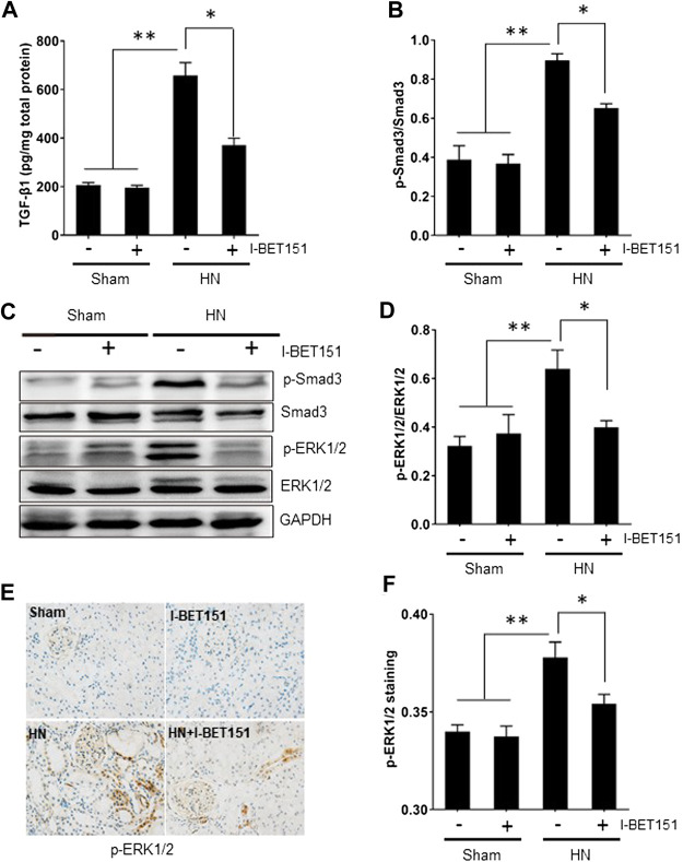
FIGURE 5.
I-BET151 abrogates the TGF-β/Smad3 signaling pathway in kidneys of HN rats. (A) Protein was extracted from kidneys and subjected to measurement of TGF-β1 by ELISA as indicated. (B) Expression levels of p-Smad3 were quantified by densitometry and normalized to Smad3. (C) The kidney tissue lysates were subjected to immunoblot analysis with specific antibodies against p-Smad3, Smad3, p- ERK1/2, ERK1/2, or GAPDH. (D) Expression levels of P-ERK1/2 were quantified by densitometry and normalized to ERK1/2. (E) Photomicrographs (original magnification, ×400) illustrate immunohistochemical staining for p-ERK1/2 in kidney tissues. (F) p-ERK1/2 staining graphic presentation of quantitative data. Data are represented as the mean ± SEM. *p < 0.05; **p < 0.01.

