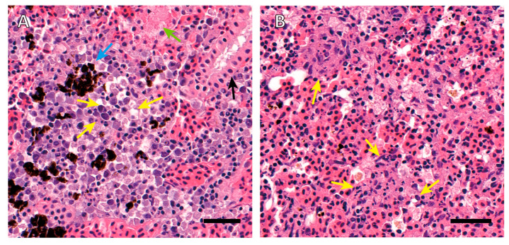
Figure 1.
Histopathology of kidney and spleen of a moribund rainbow trout with Janthinobacterium tructae strain SNU WT3 infection. (A) Histopathology of the kidney showed hyperemia and melanomacrophage accumulation in the interstitium (blue arrow). Karyolysis, pyknosis, karyorrhexis, and hydropic and vacuolar degeneration of interstitial hematopoietic cells can also be observed (yellow arrows). Epithelial cell pyknosis (black arrow) and eosinophilic droplet accumulation (green arrow) existed in renal tubules. (B) Histopathology of the spleen showed a wide range of necrotizing hematopoietic cells with eosinophilic color change. Karyolysis, pyknosis, and karyorrhexis can also be observed (yellow arrows). Slides were stained with hematoxylin and eosin. Scale bars = 40 μm.

