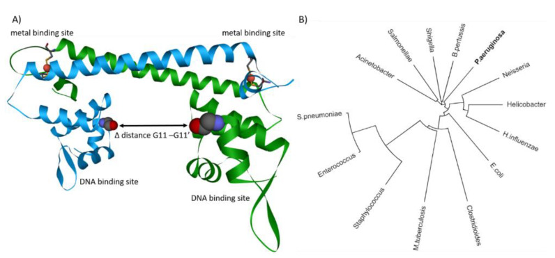
Figure 3.
(A) CueR dimer with complexed Cu(I). The two metal binding sites are on top composed of two cysteine residues. The DNA binding sites are at the bottom acting as a hinge. Upon copper binding, the distance between the two hinges is reduced, resembling a clamp, which closes upon copper binding. The difference of the distance between active and repressed state was calculated between the two G11 residues. (B) Phylogenetic tree of P. aeruginosa cueR homologs and merR family members found in pathogenic bacteria. Calculated with Clustal Omega and illustrated with iTOL [133,134].

