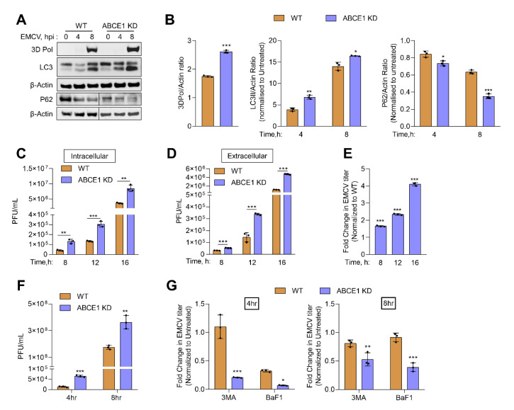
Figure 5.
Effect of ABCE1 on autophagy during EMCV infection. HT1080 WT and ABCE1 KD cells were infected with EMCV (MOI of 1.0) and at indicated times (A) conversion of unconjugated LC3-I to lipidated LC3-II, degradation of p62, and accumulation of viral protein 3D Pol were monitored on immunoblots and normalized to β-actin levels. (B) The band intensity was calculated using Image J software, and the ratio of LC3-II/β-actin, p62/β-actin, or 3D Pol//β-actin was determined and the levels were compared to WT cells. (C) Intracellular and (D) extracellular titer of EMCV were determined by a plaque assay, and (E) the fold change in EMCV titers in supernatants of ABCE1 KD cells was compared to WT cells. Results are representative of three independent experiments. (F) EMCV titers in WT and ABCE1 KD cells 4 h or 8 h post infection. (G) WT and ABCE1 KD cells were left untreated or were pretreated with 3-MA (5 mM) or bafilomycin A1 (100 nM) 1 h prior to infection with EMCV (MOI of 1.0), and viral titers in the supernatant were determined by a plaque assay. The fold change in the viral yield in WT and ABCE1 KD cells treated with either 3-MA or bafilomycin A1 was compared to untreated samples. Data represent mean ± SD performed in triplicate. WT: wild-type; * p < 0.05; ** p < 0.01; *** p < 0.001.

