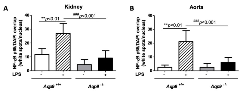
Figure 6.
Immunofluorescence evaluation of NF-κB p65 in cell nuclei. The number and intensity of white spots corresponding to the DAPI/p65 overlap in cell nuclei was evaluated by the ImageJ-win32 software using confocal microscopy images of kidneys (A) and aortas (B) harvested from Aqp9+/+ (WT) or Aqp9−/− (KO) mice sacrificed 6 h after the injection of saline solution (control) or LPS (40 mg/kg) (4 animals/group) (see Materials and Methods for details). In WT mice, LPS treatment induced a significant increase of the NF-κB p65 extent compared to WT control mice (** p < 0.01 Aqp9+/+ vs. Aqp9+/++LPS). No significant increase of the intranuclear level of NF-κB p65 was seen in the kidney and aorta of KO mice that were treated with LPS as compared to the KO control animals (### p < 0.001).

