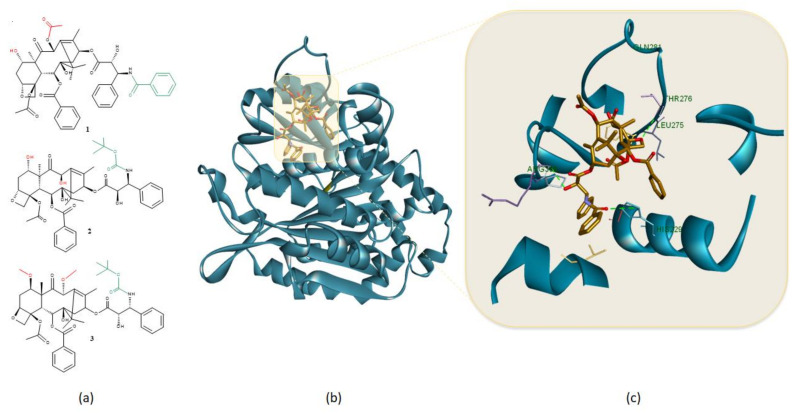
Figure 3.
(a) Chemical structures of (1) paclitaxel, (2) docetaxel, and (3) cabazitaxel, major structural differences are highlighted in green while alkylation/acylation of OH groups is highlighted in red; (b) 3D structure of stabilized microtubule chain A (blue) (PDB ID: 5SYF) in complex with taxol (gold) and (c) important HBs (green dotted lines) formed by the taxane structure within the β-tubulin binding site. Protein-ligand representation was achieved using Biovia Discovery Studio 4.1 (Dassault Systems).

