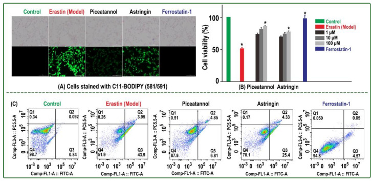
Figure 2.
Inhibitory effect of Fer-1, piceatannol, and astringin on erastin-induced ferroptosis in bmMSCs: (A) C11-BODIPY assay; (B) CCK-8 assay; the control group was cultured in medium only, while the model group was treated with erastin. The sample group was damaged by erastin and then treated with 1, 10, and 100 μM piceatannol or astringin. The positive control group was damaged by erastin and then treated with Fer-1. Each value is expressed as the mean ± SD, n = 3; *, p < 0.05, significant difference vs the model group. (C) Flow cytometry assay; the assay was conducted to distinguish live cells (Q4), necrotic cells (Q1), early apoptotic cells (Q3), and late apoptotic cells (Q2). The experiment was performed with three different batches of cells, and each batch was tested in triplicate.

