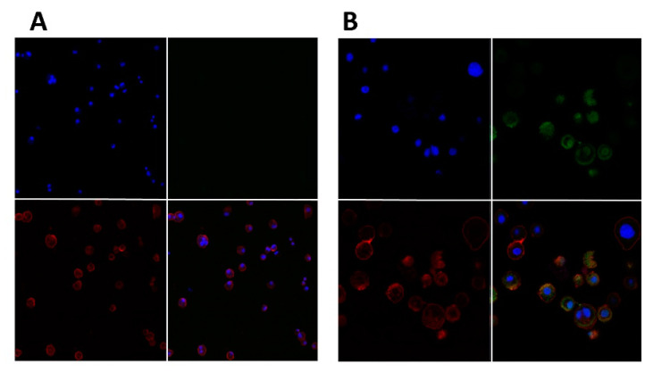
Figure 7.
Confocal microscopy images of hybrid cationic CHL NPs uptake by DCs after 1 h incubation. DCs incubated without hybrid cationic CHL NPs at 20× (A), DCs incubated with hybrid cationic CHL NPs at 63× (B). Red channel for WGA TR: cell membrane, blue channel for DAPI: nucleus, and green channel for FITC-BSA: hybrid cationic CHL NPs incorporating FITC-BSA.

