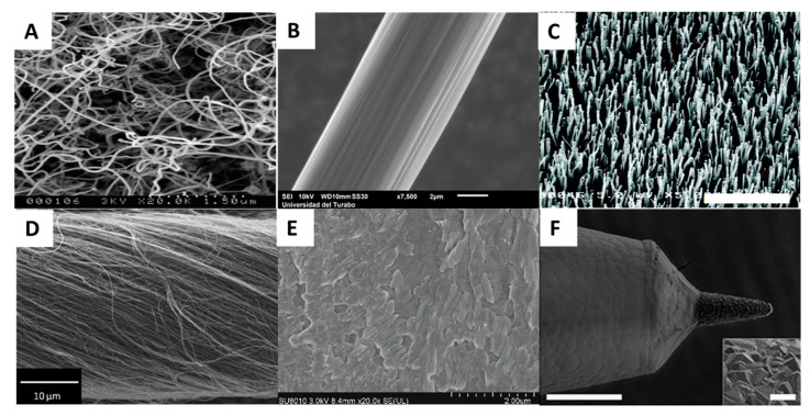
Figure 2.
SEM pictures of (A) a randomly oriented carbon nanotube; the scale bar is 1.5 µm. Adapted with permission from [91]. (B) Exposed area of carbon fiber microelectrode; the scale bar is 2 µm. Adapted with permission from [92]. (C) As-grown vertically aligned carbon nanofibers; the scale bar is 6 µm. Adapted with permission from [93]. (D) 20° twisted carbon nanotube yarn microelectrode; the scale bar is 10 µm. Adapted with permission from [82]. (E) Reduced-graphene oxide on top of a platinum electrode; the scale bar is 2 µm. Adapted with permission from [39]. (F) boron-doped diamond tip and parylene insulation; the scale bar is 100 µm. The scale bar of the inset picture is 10 µm. Adapted with permission from [54].

