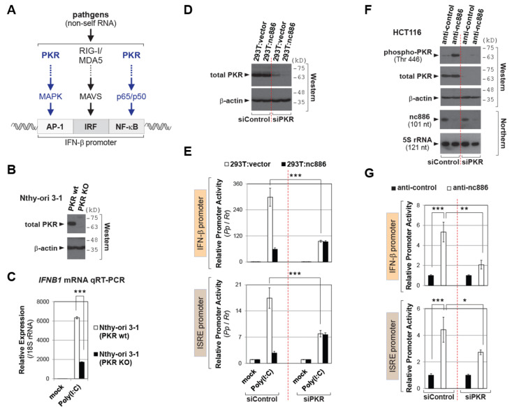
Figure 2.
nc886 suppresses the IFN promoter via inhibiting PKR. (A) Image showing the IFN-β promoter as well as regulatory factors and pathways. (B) Western blot of PKR and β-actin as a loading control. (C) qRT-PCR as described in Figure 1C. 2−ΔΔCt values were relative to the mock transfected PKR wt sample. (D) Western blot of PKR and β-actin as in panel B. (E) Luciferase assays for the reporter plasmid indicated on the left. Initial siRNA transfection (0 h); transfection of luciferase plasmids at 12 h, cell harvest and assays at 36 h. All other information is the same as Figure 1D. (F) Western and Northern blot of indicated proteins and RNAs, at 24 h after co-transfection of indicated siRNAs and anti-oligos. (G) Luciferase assays, as described in panel E and F. Luciferase plasmids were co-transfected with siRNAs and anti-oligos. Three asterisks designate a p-value less than 0.001, respectively.

