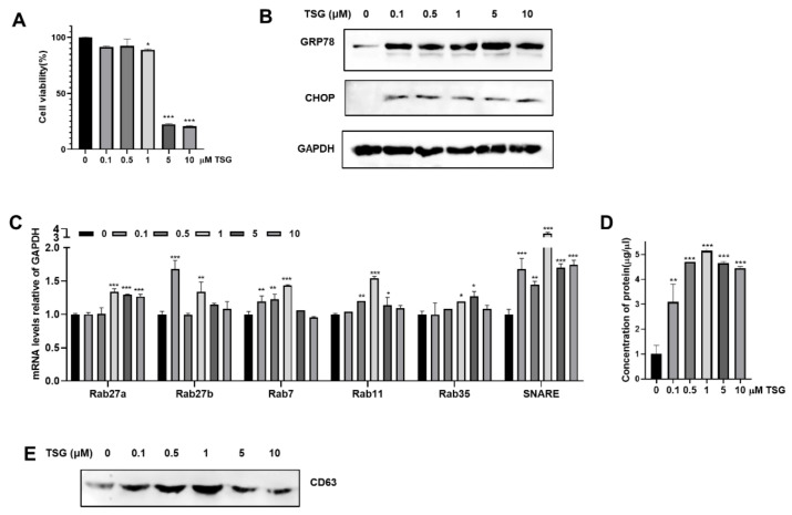
Figure 1.
Induction of ER stress by TSG stimulates EV release in WJ-MSCs. (A–C) WJ-MSCs were treated with increasing doses of TSG (0, 0.1, 0.5, 1, 5, 10 μM) for 24 h. (A) Cell viability was assessed by MTT assay. (B) The expression of GRP78 and CHOP in cell lysates (40 μg) was measured by Western blot analysis. (C) Gene expressions of Rab27a, Rab27b, Rab7, Rab11, Rab35, and SNARE in cell lysates was detected by qRT-PCR. (D,E) EVs derived from TSG treated WJ-MSCs were isolated by standard ultracentrifugation. (D) Protein concentration of EVs was measured by BCA assay. (E) Expression of CD63 EV marker in same volume (15 μL) of EV fraction was assessed by Western blot. Data are represented as the means ± S.D. of three independent experiments (* p < 0.05, ** p < 0.01, *** p < 0.001).

