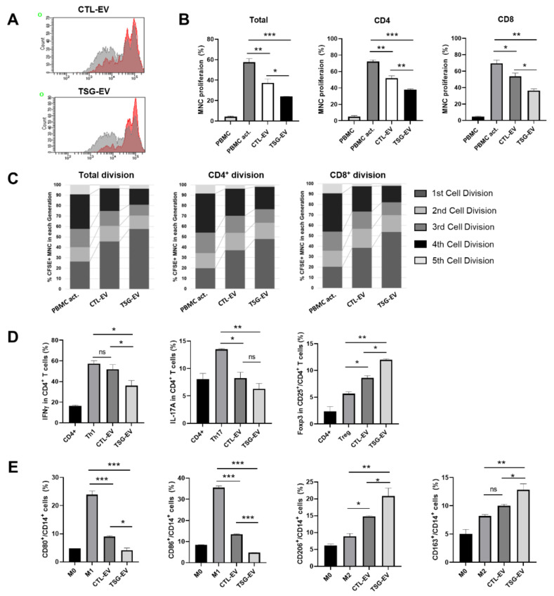
Figure 3.
EVs from TSG-primed WJ-MSCs regulate T cell proliferation and T helper (Th) cell differentiation and macrophage polarization. (A–C) CTL-EV and TSG-EV were co-cultured with CFSE-labeled PBMCs stimulated by anti-CD3/28 beads and IL-2. After 6 days, proliferation of T cell was measured by flow cytometry analysis. (A) Representative histogram of total T cells proliferations. The flow cytometry histograms show representative analyzes from three independent experiments. (B) Quantitative analysis of proliferation rates of the total T, CD4+ T, and CD8+ T cells. (C) Cell division in the five individual parts was tracked by the fluorescence profile of CFSE-labeled cells. (D) CD4+ T cells were incubated with specific lineage-driving cytokines with or without CTL-EV and TSG-EV in the presence of anti-CD3/CD28 beads and IL-2 for 5 days. The percentage of CD4+IFNγ+Th1, CD4+IL-17A+Th17, CD4+CD25+Foxp3+Treg cell was analyzed by flow cytometry and quantified. (E) CD14+ monocytes were differentiated into macrophage by GM-CSF or M-CSF for 6 days. Macrophages were activated with either the M1 cytokines (M0-GM; IFNγ + LPS) or M2 cytokines (M0-M; IL-4 + IL-13) for 48 h, respectively. Expression of M1 (CD80, CD86) and M2 (CD206, CD163) macrophage surface markers was analyzed by flow cytometry and quantified. The data are shown as the mean ± S.D. of three independent experiments (* p < 0.05, ** p < 0.01, *** p < 0.001).

