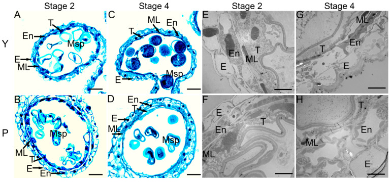
Figure 3.
Observation of transverse sections (A–D) and transmission electron microscope (TEM) (E–H) of different stages in male sterile line (P) and its maintainer line (Y). (A–D), stained with safranin O-fast green. Stage 2 (Early uninucleate stage): (A,B,E,F); Stage 4 (Binucleate stage): (C,D,G,H). E, En, ML, T, and Msp indicate the epidermis, the endothecium, the middle layer, the tapetum, and the microspore, respectively. Bars: 50 µm (A–D); 10 µm (E–H).

