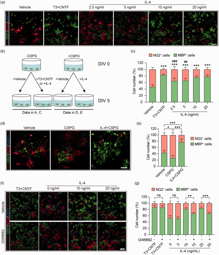
Figure 4.
IL-4 directly improves primary OPC differentiation into mature oligodendrocytes in a PPARγ-dependent manner. (a) Primary OPC cultures were treated with vehicle (PBS), T3 (50 ng/mL) + CNTF (10 ng/mL) or escalating concentrations of IL-4 for five days. Cells were triple-stained with MBP (green), NG2 (red), and DAPI (blue). Scale bar = 50 μm. (b) Schematic illustration of the experimental design in a, c, d, and e. (c) Quantification of MBP+ cells and NG2+ cells (as percentages of total cells) in panel A. ***p< 0.001 versus vehicle, ###p< 0.001 versus T3 + CNTF by one-way ANOVA and Bonferroni post hoc. (d) Primary OPC cultures were treated with vehicle (PBS), CSPG (50 ug/mL), or CSPG + IL-4 (20 ng/mL) for five days, and then immunostained for MBP (green) and NG2 (red). Nuclei were labeled with DAPI (blue). Scale bar = 50 μm. (e) Quantification of MBP+ cells and NG2+ cells as percentages of total cells. *p< 0.05, ***p< 0.001 by one-way ANOVA and Bonferroni post hoc. (f) Primary OPC cultures were treated with vehicle (DMSO) or GW9552, a PPARγ antagonist, followed by T3+CNTF and two concentrations of IL-4 for five days. Cells were then immunostained for MBP (green) and NG2 (red), and nuclei were labeled with DAPI (blue). Scale bar = 50 μm. (g) Quantification of MBP+ cells and NG2+ cells as percentages of total cells. **p< 0.01, ***p< 0.001, ns: no significance by t-test or Mann–Whitney U test. Six fields from three independent experiments were quantified.

