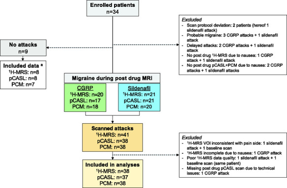
Figure 3.

Flowchart of MRI data acquisition.
aFor no attack data: three 1H-MRS, two pCASL scans and three PCM scans were acquired after sildenafil; one 1H-MRS and one pCASL scan, from the same patient, and one baseline pCASL scan were excluded due to poor quality; two PCM scan data (incl. baseline) were excluded due to insufficient flow estimate from arteries.
CGRP: calcitonin gene-related peptide; 1H-MRS: proton magnetic resonance spectroscopy; pCASL: pseudo-continuous arterial spin labeling; PCM: phase-contrast mapping; VOI: volume-of-interest; n: number.
