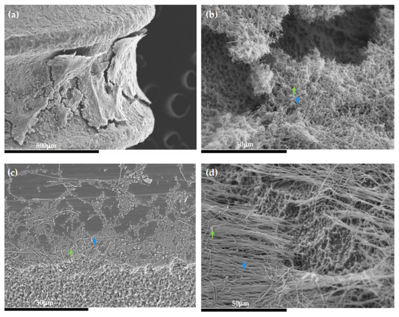
Figure 7.
Scanning electron microscopy (SEM) images of the 96-h biofilms over the Straumann® Tissue Level Standard implant. (a) View of the biofilm formation between the implant threads; (b–d) presence of spindle-shaped rods forming three-dimensional structures, which was recognized as F. nucleatum (blue arrows), with short streptococcal chains adhering, which was identified as S. oralis (green arrows), surrounded by a dense extracellular matrix covering the entire surface.

