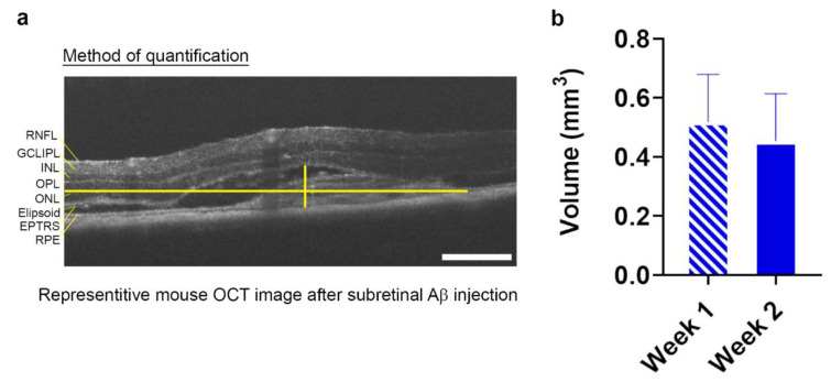
Figure 2.
Quantification of Aβ-induced GA-like lesions in living mouse retinae. Optical coherence tomography (OCT) scans of mice subretinally injected with 625 nM human oligomeric Aβ1-42 revealed the presence of a discernable focal lesion. (a) The maximal height and width of the lesion was measured using the caliper tool function as shown in the sample OCT image and (b) presented as volumetric measurements at 1 and 2 week post-injection. The average lesion volume measured 0.52 mm3 ± 0.12 SEM at 1 week and 0.45 mm3 ± 0.16 SEM at 2 weeks. n = 7 mice (p = 0.78) Two-tailed Student’s t-test. No significant differences in lesion sizes were observed between weeks 1 and 2.

