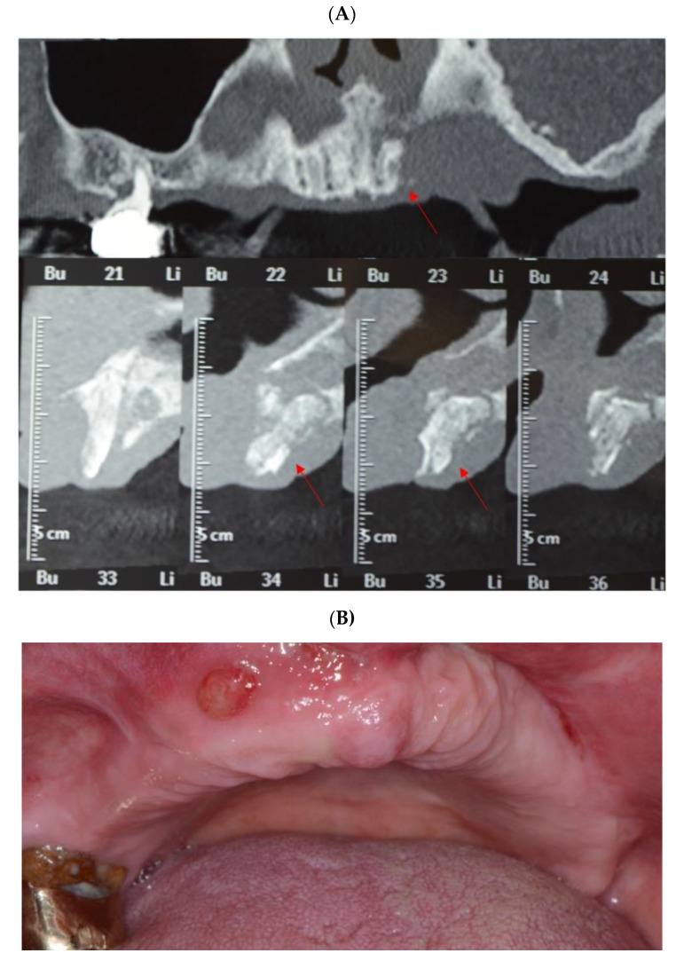
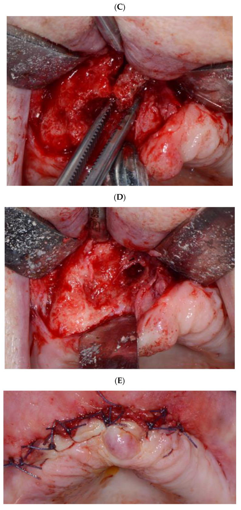
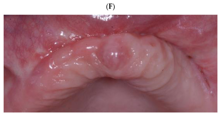
Figure 3.
Clinical case of MRONJ localized in the upper maxilla. (A) Cone beam computed tomography (CBCT) images, frontal and sagittal views, showing maxillary bone sequestration (red arrows indicate the bone sequestrum). (B) Intraoral clinical view showing the presence of fistula, which demonstrates the presence of infection. (C) Necrotic bone sequestrum removal, after the opening of the surgical flap (mid-crestal incision on the alveolar crest of the edentulous area). (D) The necrotic bone was completely removed until reaching the healthy bone tissue; bone curettage and osteoplasty were performed until vital bone was clinically observed. (E) Primary closure via periosteal releasing incisions using absorbable suture. (F) Follow-up after 3 months from the surgical intervention showing a complete tissue healing.


