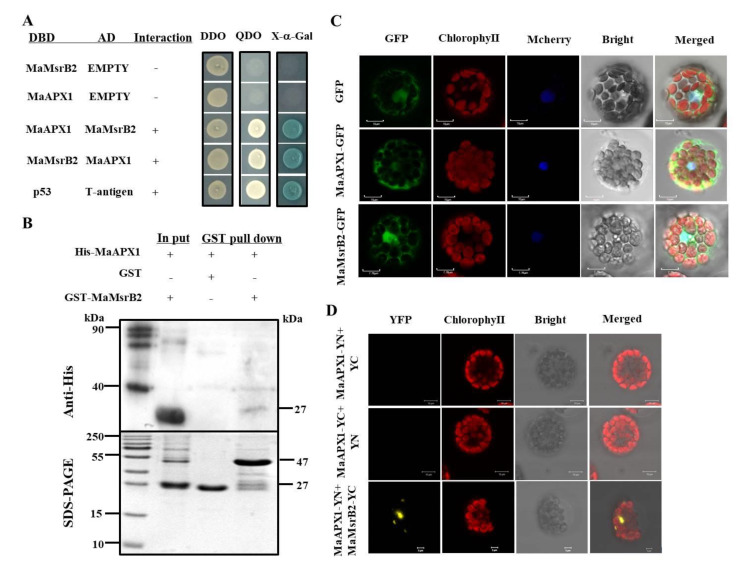
Figure 3.
MaAPX1 interacts physically with MaMsrB2. (A) Interaction between MaAPX1 and MaMsrB2 in the yeast two-hybrid (Y2H) assay. (B) Interaction between MaAPX1 and MaMsrB2 in GST pull-down assay. The molecular weight of MaAPX1-His and MaMsrB2 is about 27-kDa and 47-kDa, respectively. (C) Subcellular localization of MaAPX1 and MaMsrB2. A green signal indicates green fluorescent protein (GFP) fluorescence; A red signal indicates chlorophyll auto fluorescence; The merged images represent a digital combination of chlorophyll auto fluorescence and GFP fluorescent images. (D) Interaction between MaAPX1 and MaMsrB2 in the bimolecular fluorescence complementation (BiFC) assay. A yellow signal indicates yellow fluorescent protein (YFP) fluorescence; a red signal indicates chlorophyll autofluorescence; the merged images represent a digital combination of the chlorophyll autofluorescence and YFP fluorescent images.

