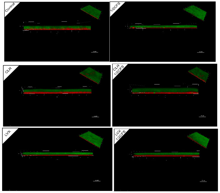
Figure 7.
CLSM images of a 1/2 × MIC dilution of CFS, 1 × MIC dilutions of CLR and LVX alone, or the same dilutions of CFS and antibiotic combinations on H. pylori mature biofilm structure. All cells stained with SYTO 9 show green fluorescence, and dead cells stained with propidium iodide (PI) show red fluorescence. The bottom layer of the biofilm in the main view is close to the NC membrane surface. The front view shows the horizontal observations along the x-y plane, and the scale bar is 20.00 μm.

