Figure 1.

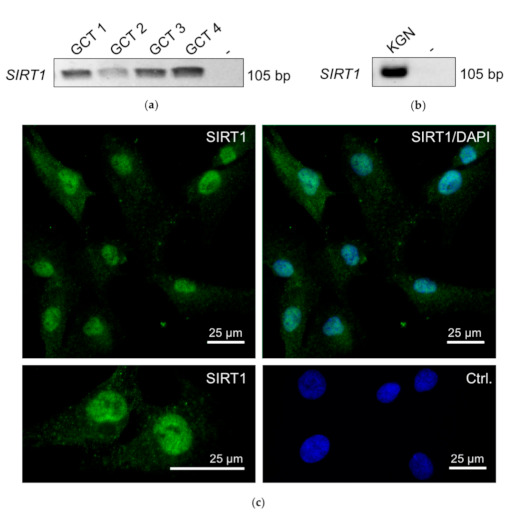
RT-PCR revealed SIRT1 mRNA (single band of 105 bp) in granulosa cell tumor (GCT) samples from four patients (a). Non template (-) control was negative (instead of cDNA, H2O was used). (b) SIRT1 was also detected in KGNs (RT-PCR). (c) Micrograph of immunofluorescence staining of SIRT1 in KGN. Upper left: SIRT1 staining (green) was found in the nuclei; upper right micrograph: SIRT1 staining merged with DAPI (blue). Left lower panel: higher magnification of the SIRT1 staining. Left lower panel: control: omission of the first antibody merged with DAPI. Bars represent 25 µm.
