Figure 4.

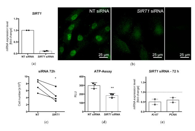
qPCR analysis showed decreased mRNA levels of SIRT1 after siRNA treatment for 72 h (a). Immunofluorescence of KGN after siRNA treatment (72 h): while in nontarget (NT) siRNA-treated cells, prominent SIRT1 staining (green) was observed mainly in the nucleus, silencing of SIRT1 strongly reduced cellular staining (b). Ctrl., control experiment (omission of first antibody). Cell numbers (n = 4) and ATP levels (n = 3) were significantly reduced after siRNA silencing (c,d) (* p < 0.05, ** p < 0.005, t-test). The levels of the proliferation markers Ki-67 and PCNA were decreased in SIRT1-siRNA-treated KGN, as shown by qPCR analysis (e) (means and SEMs; n = 2).
