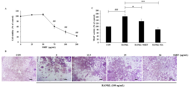
Figure 2.
SMEF suppresses osteoclastogenesis in vitro. (A) RAW264.7 cells cultured with SMEF in concentration range of 0–200 μg/mL for 48 h in 96-well plates. Cell viability was determined by an MTT assay. ## p < 0.001 compared with untreated control. (B) RAW264.7 cells were coincubated with SMEF (0–50 μg/mL) and RANKL (100 ng/mL) for 6 days and then stained using a leukocyte acid phosphatase (TRAP) kit. TRAP-positive multinucleated osteoclasts were visualized in 100× magnification under light microphotography. Scale bars, 100.2 μm. (C) RAW264.7 cells were co-incubated with SMEF (50 μg/mL) and RANKL (100 ng/mL) for 6 day and then measured using the TRAP solution assay. RA (100 μM) was used as a positive control and TRAP activity was expressed as % of control. The data is presented as the mean ± SD of three independent experiments. ## p < 0.001, compared with control (CON); * p < 0.01, ** p < 0.001 compared with RANKL control.

