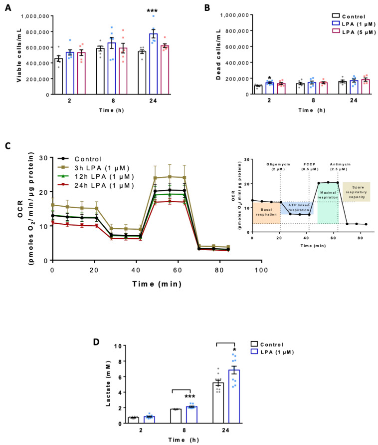
Figure 1.
Effect of lysophosphatidic acid (LPA) on cell viability, mitochondrial respiration and lactate content of BV-2 microglia. The numbers of (A) viable and (B) dead cells were determined by Guava ViaCount analysis in the absence and presence of LPA. (C) Oxygen consumption rate (OCR) in the absence and presence of 1µM LPA for the indicated times was detected using the XF Cell Mito Stress Test. Cells were treated with 2 μM oligomycin, 0.5 μM FCCP, and 2.5 μM antimycin A in XF assay medium to assess fundamental parameters of mitochondrial function (inset). (D) Lactate content in the supernatants of LPA (1 µM) treated cells was measured by EnzyChrom™ Glycolysis Assay Kit and compared to their appropriate controls. Bar graph represents mean values ± SEM of 3 independent experiments; (* p < 0.05, *** p < 0.001 compared to control; one-way ANOVA with Bonferroni correction).

