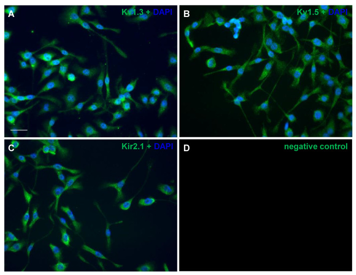
Figure 1.
Immunolabeling of potassium channels in cultured BV2 microglial cells. The cells were stained with antibodies targeting Kv1.3 (A), Kv1.5 (B), Kir2.1 (C) and the negative control with Alexa 568 (D). The coverslips were mounted using the Prolong antifade with DAPI in blue. The channel was pseudo-colored in green for improved visualization. The images are representative of three independent experiments. Scale bar: 40 µm.

