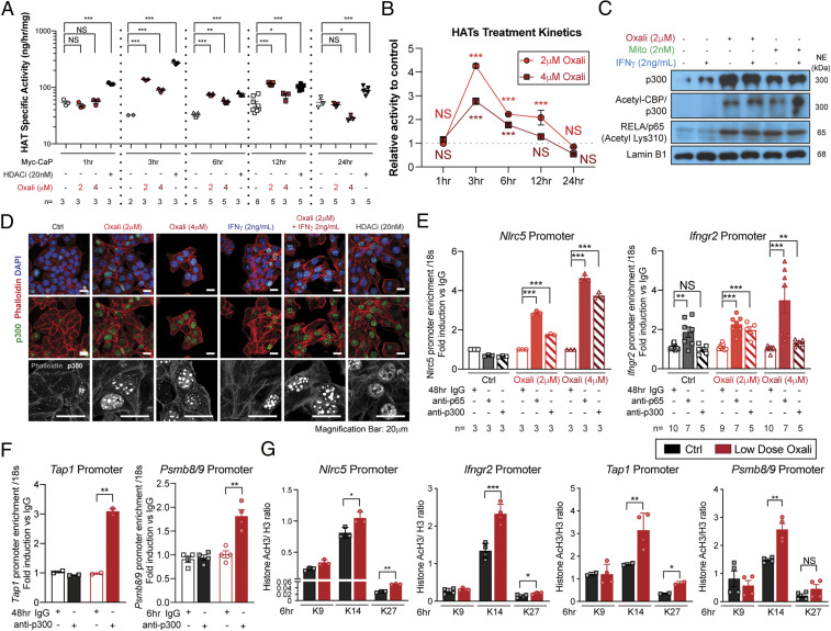
Fig. 3.
Oxaliplatin and mitoxantrone stimulate HAT activity and nuclear localization. (A and B) Myc-CaP cells incubated with Oxali (2 or 4 μM) or the HDACi panobinostat (LBH589; 20 nM) for the indicated times were lysed and analyzed for HAT activity using H3 as a substrate. (C) Nuclear extracts of Myc-CaP cells treated with Oxali, Mito, and/or IFNγ were IB analyzed for p300, acetylated-CBP/p300, acetylated-RELA/p65 (lysine K310), and lamin B1 (loading control). (D) Myc-CaP cells treated as indicated for 12 h were stained with anti-p300 (green) and phalloidin (red; actin cables). Nuclei were counterstained with DAPI (blue). (Scale bar: 20 μm.) (E–G) Untreated and Oxali-treated Myc-CaP cells were subjected to ChIP analysis with control IgG and antibodies to p65/RelA, p300 as indicated (E and F) or acetylated H3 (lysine K9, K14, and K27) (G). Precipitation of the indicated promoter regions was determined by PCR. Two-sided t test (means ± SEM), and Mann–Whitney test (median) were used to determine significance. *P < 0.05; **P < 0.01; ***P < 0.001; NS, not significant. Specific n values are shown in A and E; each experiment includes at least three biological replicates.

