Fig. 1.
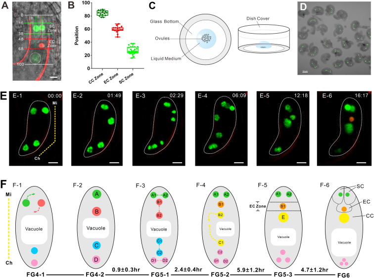
The nucleus migrates to a specific position before cell specialization in the FG. (A) FGR7.0 is a triple marker line with RFP in the egg cell nucleus, bright GFP in synergid cell nuclei, and weak YFP in the central cell. In mature FGs, the nuclear positions of synergid cells are in the SC zone, the nuclear position of the egg cell is in the EC zone, and the nuclear position of the central cell is in the CC zone. The numbers are the relative positions of FGs. (Scale bar: 10 µm.) (B) Relative positions of the SC, EC, and CC zones. The data are calculated from ovules observed (n = 15). (C) Diagram of the in vitro ovule culture system. A Nitsch medium droplet containing ovules was added to the glass bottom of a cell culture dish. (Left) Top view. (Right) Front view. (D) A group of ovules at FG4 collected for culture. (Scale bar: 50 µm.) (E) Time lapse of NPCI FG development from FG4 to FG6. The yellow dotted line indicates the micropylar (Mi) end and the chalazal (Ch) end of the FG in E-1. The numbers in the right corner of each image are the culture times (h: min). In E-6, FG exhibits an egg cell. The whole process can be seen in Movie S1. (Scale bar: 10 µm.) (F) Diagram of FG development corresponding to E. The corresponding developmental stages and duration time (h) are indicated below the images. The duration time (mean ± SD) was calculated from ovules observed (n = 11). The yellow dotted line indicates the Mi end and the Ch end of FG in F-1. The dotted arrows in F-1 show the direction of nuclear migration. In F-2, the four nuclei are arranged along the micropylar-chalaza axis and labeled as nuclei A, B, C, and D. In F-3, four nuclei divide to give rise to eight nuclei. Nuclei A1 and A2 are derived from nucleus A, nuclei B1 and B2 are derived from nucleus B, nuclei C1 and C2 are derived from nucleus C, and nuclei D1 and D2 are derived from nucleus D. In F-4, nuclei B2 and C1 move to each other. The dotted arrows show the direction of nuclear migration. In F-5, nuclei B2 and C1 fuse to form one large nucleus, E. Nucleus B1 is located in the EC zone. In F-6, the nucleus B1 shows an egg cell identity. Nuclei A1 and A2 located in the SC zone show synergid cell identity.

