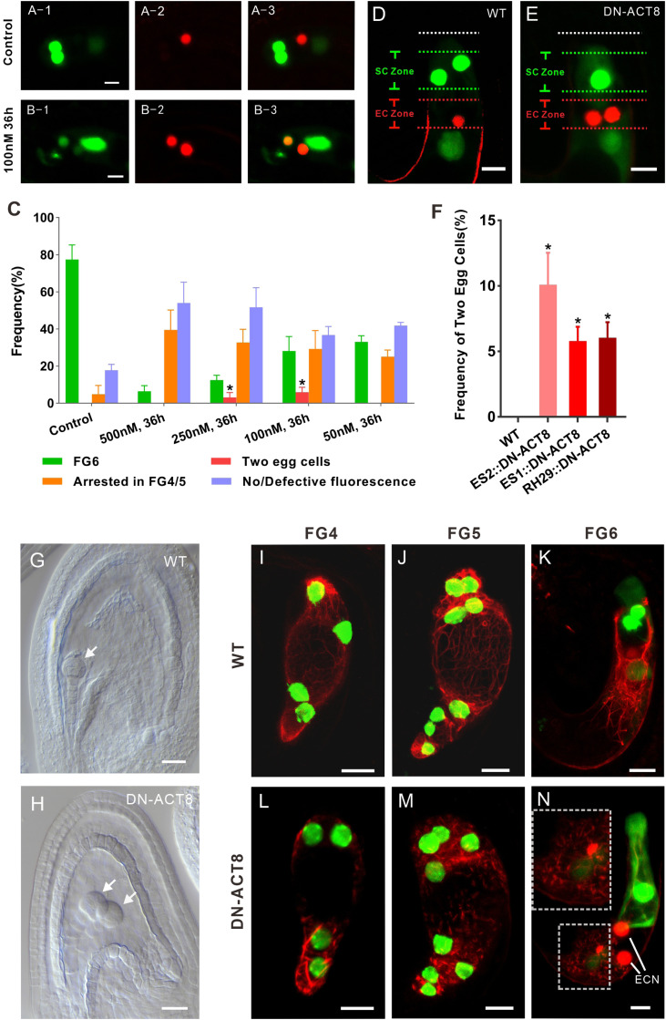
Fig. 2.
Disruption of the nuclear position resets the cell identity of the egg apparatus. (A) In the control, there is only one egg cell. (B) Treatment with the inhibitor of actin polymerization LATB results in two egg cells. (C) LATB affects FG development and egg cell specialization. Ovules treated with a high concentration of LATB (500 nM) and FGs seldom develop to FG6, with most FGs arresting in FG4 or FG5 (FG4/5) or losing fluorescence. Treatment with 200 nM or 100 nM LATB, but not with 50 nM LATB, results in two egg cells. Most FGs develop to FG6 in the absence of LATB. At least three biological replicates were performed for each treatment, containing 15 to 26 ovules at FG4-1. The total number of ovules observed was 79, 75, 106, 91, and 87 for the control and 500 nM, 250 nM, 100 nM, and 50 nM LATB treatments, respectively. The data are mean ± SD. Significant differences (*P < 0.05) were determined using the paired t test. (D) In WT, FGs contain two synergid cells and one egg cell at FG6 stage. (E) In transformed plants expressing DN-ACT8, FGs contain one synergid cell and two egg cells at FG6 stage. (F) Frequency of FGs exhibiting two egg cells in transformed plants expressing DN-ACT8. DN-ACT8 is expressed under the control of three FG-specific promoters—ES1, ES2, and RH29—to disturb actin filaments. The data (mean ± SD) were counted from FG at FG6 stage of WT (n = 245), ES1:: DN-ACT8 (n = 366), ES2:: DN-ACT8 (n = 389), and RH29:: DN-ACT8 (n = 412). Significant differences (*P < 0.05) were determined using the paired t test. (G) In WT, there is a globular embryo at 3 d after fertilization. (Scale bar: 20 µm.) (H) In the DN-ACT8 line, there are two globular embryos at 3 d after fertilization. (Scale bar: 20 µm.) (I–K) In WT, the actin cable labeled by Lifeact-Tag-RFP can be clearly seen in FGs at FG4-FG6. (L–N) In proES2::DN-ACT8 plants, the actin filaments became much shorter and generated aggregates in FG at FG4-FG6. In L, the FG at FG6 has two egg cells. The upper-left corner of the rectangle is an enlarged view of the lower-left of the rectangle, which shows that actin filaments are disrupted. (Scale bar: 10 µm.)

