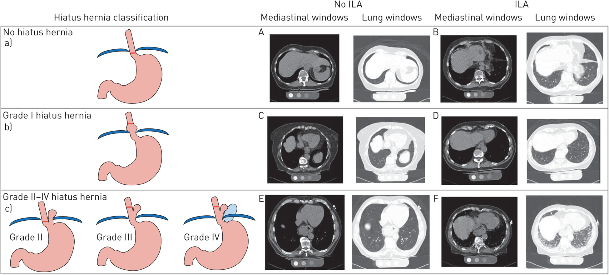
FIGURE 1.

Hiatus hernia and interstitial lung abnormality (ILA) classification. Graphical representation of a) normal thoracoabdominal anatomy (no hiatus hernia), b) grade I hiatus hernia and c) grades II–IV hiatus hernia. Mediastinal and lung windows from axial chest computed tomography scans demonstrate hiatus hernia anatomy and parenchymal involvement respectively for participants with and without ILA (A–F). Participant A has no hiatus hernia and no ILA. Participant B has no hiatus hernia and subpleural ILA with fibrosis. Participant C has a grade I hiatus hernia and no ILA. Participant D has a grade I hiatus hernia and subpleural ILA without fibrosis. Participant E has a grade II–IV hiatus hernia and no ILA. Participant F has a grade II–IV hiatus hernia and subpleural ILA with fibrosis.
