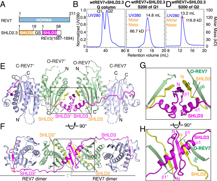
Fig. 2.
Crystal structure of SHLD2.3–REV74 complex. (A) Schematic drawing of human REV7 and SHLD2.3 fusion protein. (B) Q column purification of the complex SHLD2.3 bound to REV7 yields two peaks labeled Q1 and Q2. (C) Size exclusion S200 purification of peak labeled Q1 and measurement of a 66.7-kDa molar mass for the major peak by SEC-MALS. (D) Size exclusion S200 purification of peak labeled Q2 and measurement of a 116.9 kDa molar mass by SEC-MALS. (E and F) Two views of the overall structure of SHLD2.3–REV74 complex. C-REV7, light blue; O-REV7, green; SHLD3, magenta; SHLD2, yellow. (G and H) Expanded views of the boxed segment in E (see G) and F (see H) of the complex highlighting the dimeric interface whereby SHLD2 β1–SHLD3 β1–REV7 β6 segments form a pair of β-sheets.

