Fig. 7.
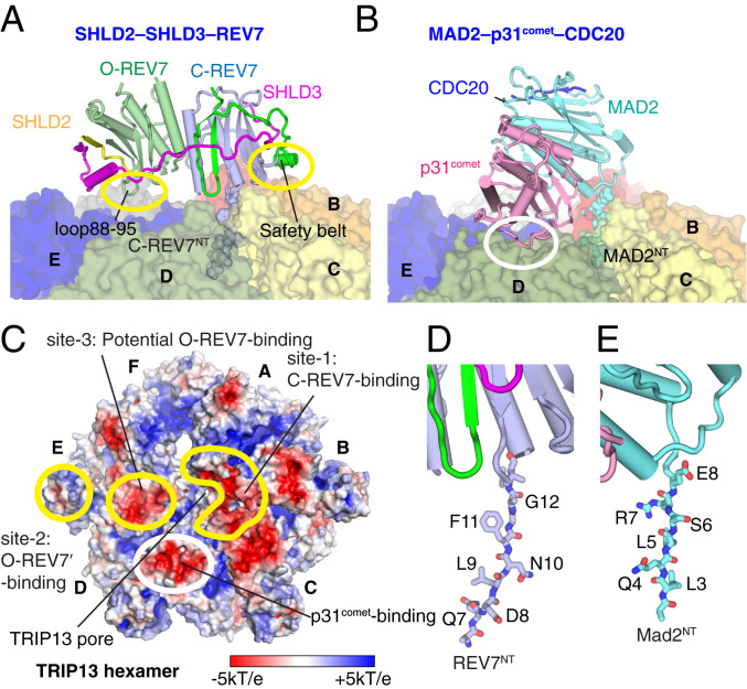
Comparison of structures of SHLD2.3–REV74–TRIP13 and MAD2–p31comet–CDC20–TRIP13 complexes. (A) Positioning of SHLD2.3-mediated O-REV7–C-REV7 dimer on the surface of TRIP13 following insertion of REV7NT into the central pore of TRIP13 in the SHLD2.3–REV74–TRIP13 structure. (B) Positioning of MAD2–p31comet–CDC20 on the surface of TRIP13 following insertion of MAD2NT into the central pore of TRIP13. (C) Electrostatic surface representation of the TRIP13 (surface potential at ±5 kT e−1). Intermolecular contact patch between REV7 dimer and TRIP13 are highlighted by labeled yellow circles while the intermolecular contact patch between p31comet and TRIP13 is highlighted by a white circle. Monomers D and E use the consistent acidic surface for p31comet and O-REV7 binding, respectively. The TRIP13 pore is also indicated. The (D) REV7NT and (E) MAD2NT residues inserted into the TRIP13 pore.

