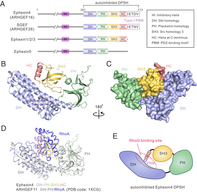
Fig. 1.
Crystal structure of Ephexin4DPSH. (A) Schematic diagrams showing the conserved domain organizations of Ephexins and SGEF. The PBM sequences of Ephexin4 and SGEF are shown. The domain color coding is consistent throughout this paper. The domain keys are also shown here. (B) Ribbon diagram representation of the Ephexin4DPSH structure. (C) Surface representation showing the overall architecture of the Ephexin4DPSH. (D) Superposition of ARHGEF11 DH-PH/RhoA (PDB ID code: 1XCG) and the Ephexin4DPSH (this study) structures. (E) Schematic representation of autoinhibited Ephexin4DPSH. The circle indicates the DH active site.

