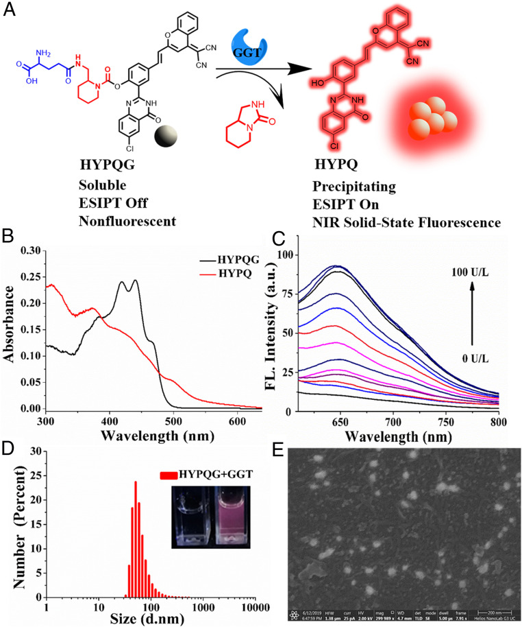
Fig. 2.
The chemical structure and photophysical properties of HYPQG. (A) The response mechanism of HYPQG with GGT that shows turn-on NIR solid-state fluorescence. (B) UV-Vis absorption spectra of HYPQG (10 μM) in DMSO and HYPQ (10 μM) in glycerol: PBS = 1:1. (C) Fluorescence emission spectra of HYPQG (5 μM) with increasing concentration of GGT (0, 1, 3, 5, 10, 15, 20, 30, 40, 60, 80, 90, and 100 U/L). λex/em = 450/650 nm. (D) Particle size distributions of HYPQG (10 μM) after reaction with GGT (150 U/L). (Inset) Photos of HYPQG (10 μM) before (Left) and after (Right) reaction with GGT (150 U/L) under UV lamp at 365-nm excitation. d, diameter. (E) SEM photos of HYPQG after reaction with GGT. HFW, horizontal field width; HV, high voltage. (Scale bar, 200 nm.)

