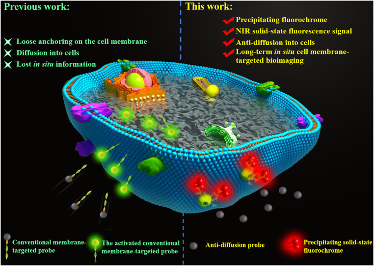
Scheme 1.
Schematic diagram illustrates the difference between conventional cell membrane-targeted bioimaging probes and our de novo anti-diffusion bioimaging probe. Traditional membrane-associated probes easily diffuse into cells, while the anti-diffusion probe releases a strong hydrophobicity and low lipophilicity fluorochrome that precipitates at the reaction sites in a manner that inhibits diffusion into the cell, hence, realizes long-term in situ cell membrane-targeted bioimaging.

