Fig. 3.
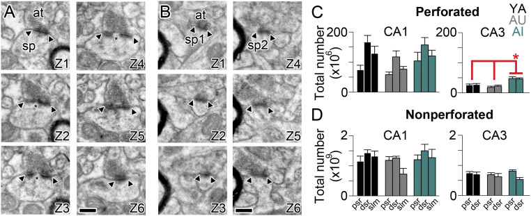
Unbiased estimate of total number of perforated and nonperforated synapses in apical dendritic domains of CA1 and CA3 hippocampus. (A) Six serial electron micrographs through a perforated axospinous synapse (Z1–Z6), with the borders of the PSD indicated by the arrowheads, and the discontinuity or perforation in the PSD indicated by an asterisk. (B) Six serial electron micrographs through two different nonperforated axospinous synapses (sp1 and sp2; Z1–Z6), with the borders of the continuous PSDs indicated by the arrowheads. In all micrographs: at, axon terminal; sp, spine. (Scale bars, 500 nm.) (C) Total number (in millions) of perforated synapses in three dendritic regions that are progressively distal to the soma/axon (dsr, distal one-third of the SR; psr, proximal one-third of the SR; slm, stratum lacunosum-moleculare) in region CA1 (Left) and CA3 (Right) of the hippocampus in YA (black), AU (gray), and AI (aqua) rats. Asterisk indicates a main effect of group attributable to AI rats having significantly more perforated synapses in CA3 pSR and dSR as compared to either YA or AU rats: F(2, 6) = 10.83, P = 0.01. (D) Same as C but for nonperforated synapses (in billions).

