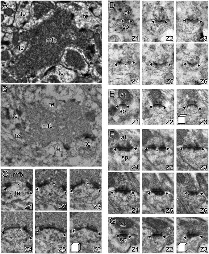
Fig. 5.
Synaptic receptor expression at excitatory synaptic subtypes onto CA1 and CA3 pyramidal neurons. (A) A conventional electron micrograph illustrating the unique morphology of synapses between granule cells in the dentate gyrus and CA3 pyramidal neurons, which are present in SL of hippocampal region CA3. The mossy fiber bouton (mfb) is densely packed with neurotransmitter vesicles and forms multiple synapses with thorny excrescences (te) on the proximal dendrites of CA3 pyramidal neurons. (B) The unique morphology of the mossy fiber–thorny excrescence (mfb and te, respectively) synaptic complexes is also present in hippocampal tissue prepared for postembedding immunogold EM using freeze substitution embedding in Lowicryl HM20. (C) A high-magnification series of electron micrographs (Z1–Z6) through a perforated synapse between an mfb and a te, immunostained for AMPARs using postembedding immunogold EM. Arrowheads denote the edges of the PSD, with the asterisk indicating the presence of a perforation or discontinuity in the PSD. (D) Serial electron micrographs of a perforated synapse in the SR (Z1–Z3), immunostained for AMPARs using postembedding immunogold EM. Arrowheads denote the edges of the PSD, with the asterisk indicating the presence of a perforation or discontinuity in the PSD. at, axon terminal; sp, dendritic spine. (E) Serial electron micrographs of a nonperforated synapse in the SR (Z1–Z3), immunostained for AMPARs using postembedding immunogold EM. Arrowheads denote the edges of the PSD. (F and G) Same as D and E, but for NMDAR-type receptor expression. (Linear dimensions of scale cubes in all micrographs are 500 nm in all axes.)

