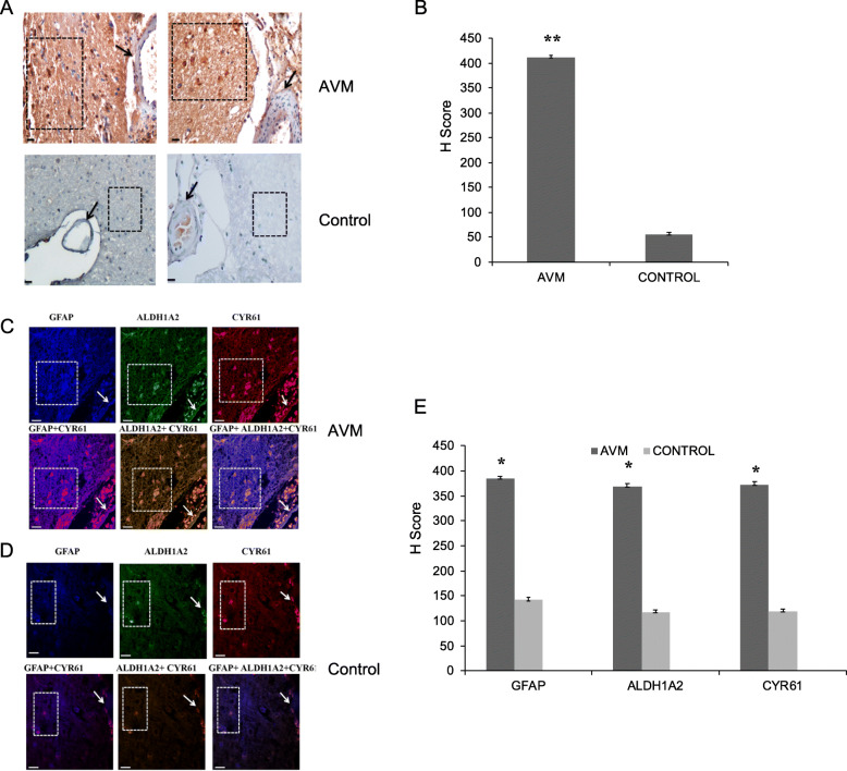
Fig. 5.
CYR61 expression analysis in cerebral AVM nidus and its neighboring astrocytes. a Representative images of immunohistochemistry analysis for CYR61 protein using antibody with cerebral AVM nidus and control tissues. The CYR61 is highly expressed in AVM brain parenchyma (box) and less expressed in AVM blood vessel (arrow). In control tissues, the CYR61 protein is not expressed in brain parenchyma (box) as well as in control blood vessel (arrow). The images were collected using × 20 magnification (scale bar, 100 μm). b The bar plot represents the H score analysis of CYR61 expression in AVM and control tissue. There is an increased expression of this protein in AVM tissues compared to control tissues (**P < 0.01), AVM (n = 10) and control (n = 10). c Photomicrograph representing triple immunofluorescence staining with GFAP (blue), ALDH1A2 (green), and CYR61 (red) in cerebral AVM tissues. Immunofluorescence staining with GFAP, ALDH1A2, and CYR61 reveals high expression of these proteins in astrocytes (box) surrounding the AVM vessel (arrow). The GFAP+ ALDH1A2 + CYR61 triple immunofluorescence staining demonstrates the co-localized expression of these three proteins in AVM-associated astrocytes. d The triple immunofluorescence staining with control tissues shows minimal expression of all the three proteins in astrocytes (box) surrounding the control blood vessel (arrow). e The bar plot represents the mean fluorescent intensity analysis of GFAP, ALDH1A2, and CYR61 in AVM and control vessels. There is an increased expression of these proteins in AVM compared to control tissues (*P < 0.05), AVM (n = 10) and control (n = 10)

