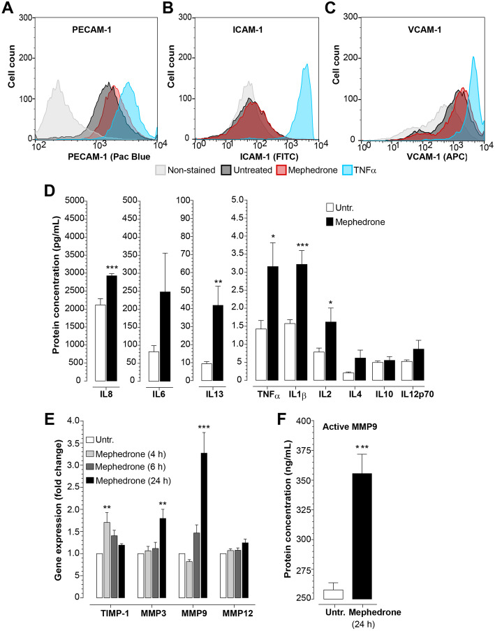
Fig. 4.
Mephedrone induces inflammatory activation of hBMVEC. a–c hBMVECs were treated with 10 μM mephedrone for 2 h and analyzed by flow cytometry. Histograms show the expression of adhesion molecules PECAM-1, ICAM-1, and VCAM-1 in response to mephedrone, which was statistically significant only for PECAM-1 at 2 h. Experiments were independently performed three times. Within each individual experimental set, primary cells from three different donors were used (n = 9). d hBMVEC were treated with 10 μM mephedrone for 24 h, and levels of cytokines were analyzed by V-PLEX assay. Levels of IL-1β, IL-2, IL-8, IL-13, and TNFα were significantly upregulated by the treatment. Data shown as the mean concentration ± SEM expressed in pg/mL. Experiments were independently performed three times. Within each individual experimental set, primary cells from three different donors were used (n = 9). e TIMP-1, MMP3, MMP9, and MMP12 mRNA expression in hBMVEC treated with 10 μM mephedrone for 4, 6, or 24 h were determined using qRT-PCR. Data shown as fold change relative to untreated control and normalized to the housekeeping gene (18S). Experiments were independently performed three times. Within each individual experimental set, primary cells from three different donors were used (n = 9). f MMP9 enzyme activity in the culture medium of endothelial cells treated with 10 μM mephedrone for 24 h were determined using Human Active MMP9 Fluorokine assay. Data shown as mean ± SEM and expressed in pg/mL of active enzyme. Experiments were independently performed three times. Within each individual experimental set, primary cells from three different donors were used (n = 9)

