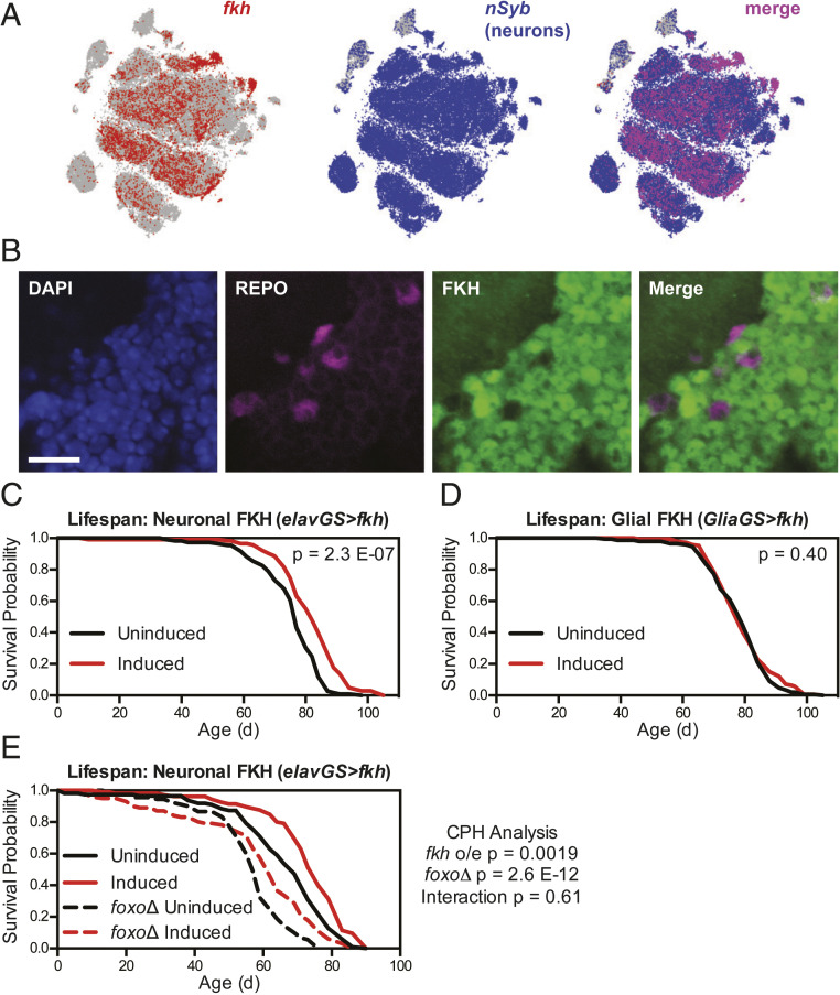
Fig. 2.
Overexpression of fkh in neurons, but not glia, extends lifespan independent of FOXO. (A) Images from the SCope database (33) show mRNA expression of fkh largely in nSyb-expressing (neuronal) cell populations in the Drosophila brain. (B) Immunofluorescence images from the cell body layer of the central brain show FKH expression in REPO-negative (neuronal) cells in wDah flies. (Scale bar, 10 μm.) (C–E) Survival curves show (C) extended lifespan for wDah;UAS-fkh/+;elav-GS/+ flies, (D) no change in lifespan for wDah;UAS-fkh/+;GSG3285-1/+ flies, and (E) extended lifespan for both wDah;UAS-fkh/+;elav-GS/+ and wDah;UAS-fkh/+;elav-GS, foxoΔ/foxoΔ flies treated with 200 μM RU-486 from 2 d of age, with no significant interaction between fkh overexpression and foxoΔ genotype. For all survival experiments, n > 85 deaths were counted per condition; P values are from either log-rank tests between groups (C and D) or Cox proportional hazards testing (E).

