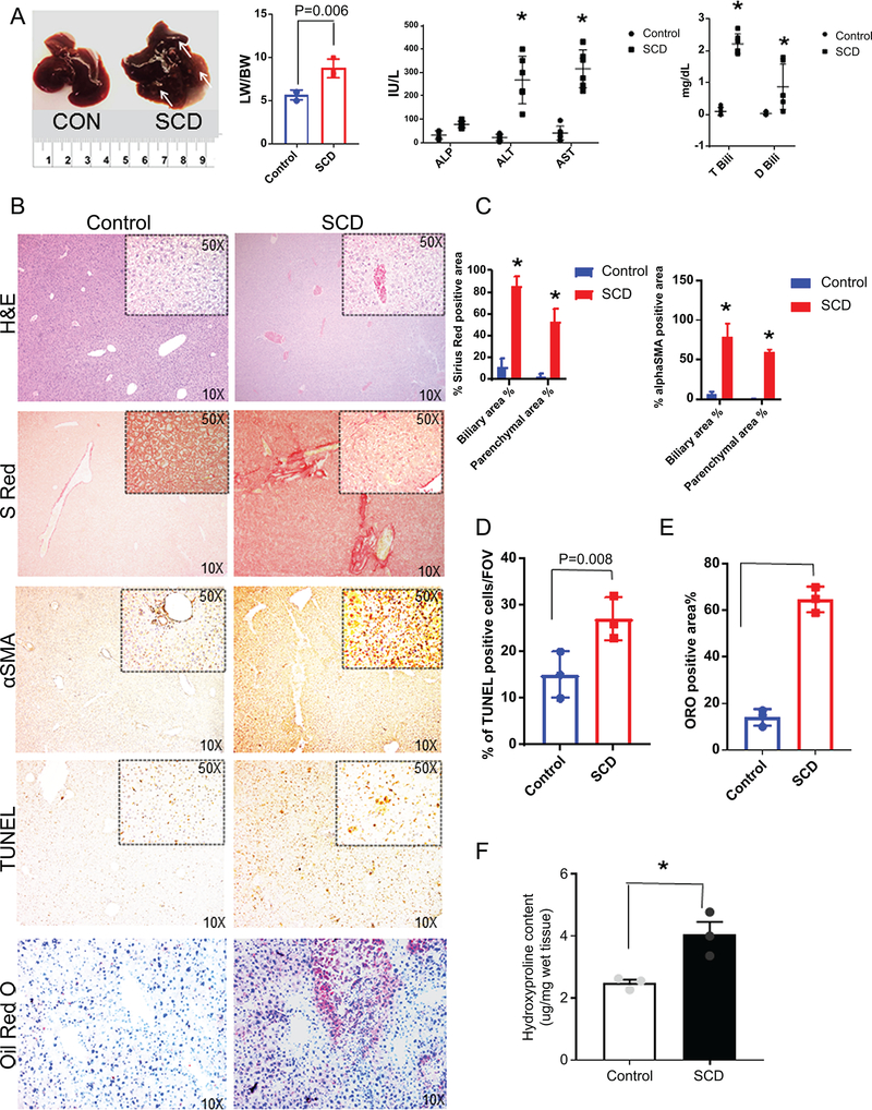
Figure 2: SCD mice have progressive liver injury and hyperbilirubinemia.
(A) Gross specimen of livers of control and SCD mice. SCD mice exhibit increased liver weight to body weight ratio. Serum biochemical analysis of liver enzymes ALT, AST and ALP in control and SCD mice. Serum analysis of direct and total bilirubin levels in control and SCD mice.(B) Immunohistochemical characterization of control and SCD mice. H&E, sirius red, αSMA, TUNEL and Oil-Red-O staining of control and SCD liver sections revealed increased injury, fibrosis, apoptosis inflammation and steatosis in SCD liver. (C) Quantification of biliary and parenchymal fibrosis as seen by Sirius red and αSMA positive staining. (D) Quantification of TUNEL positive cells in control and SCD liver. (E) Quantification of Oil-Red-O staining in control and SCD liver. (F) Hydroxyproline content in the livers from control and SCD mice. * denotes p<0.05.

