Figure 2.
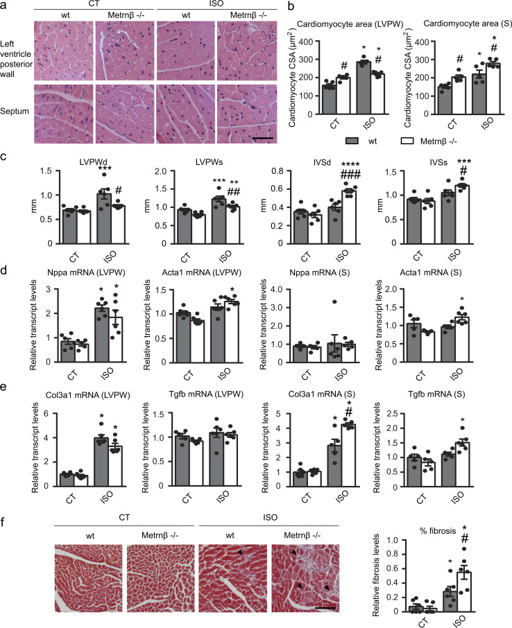
Metrnβ-null mice develop enhanced cardiac alterations in response to ISO infusion. 2-mo-old WT littermates (gray bars) and Metrnβ−/− (white bars) mice were continuously infused with ISO for 7 d to experimentally induce heart hypertrophy. (a) Representative histological sections of hearts stained with H&E were used to determine cardiomyocyte CSA. Magnification, 20×. Scale bar, 50 µm. (b) Quantification of cardiomyocyte CSA in the LVPW (P values are 0.0011, <0.0001, 0.0368, <0.0001) and theseptum (S; P values are 0.0008, 0.0047, 0.0001, 0.013). (c) IVS and LVPW in systole (s) and diastole (d) assessed by echocardiography. P values are 0.0111, 0.043, 0.0046, 0.0006, 0.0274, <0.0001, 0.0011, 0.0004, 0.0492. (d) mRNA expression of the hypertrophy marker genes Nppa and Acta1 in the LVPW and the septum (S; P values are <0.0001, 0.0089, <0.0001, 0.0008). (e) mRNA expression of the fibrosis markers Col3a1 and Tgfb in the LVPW and the septum (S; P values are <0.0001, <0.0001, 0.0018, <0.0001, 0.0122, 0.0031). (f) Determination of fibrosis in histological sections by Masson’s trichrome staining. Magnification, 20×. Scale bar, 100 µm. Arrowheads show fibrotic areas (blue; P values are 0.0238, 0.0005, 0.0479). Results are expressed as mean ± SEM; n = 6 mice/group. Data were analyzed by one-way ANOVA (*, P < 0.05; **, P < 0.001; ***, P < 0.0001, ****, P <0.00001 compared with corresponding control [CT] mice; #, P < 0.05; ##, P < 0.001; ###, P < 0.0001 compared with corresponding WT mice).

