
Figure 2: CSNK2B controls ATF6α signaling upstream of intramembrane cleavage.
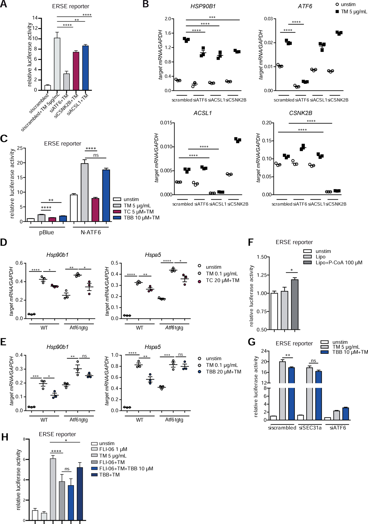
(A–B) siRNA-mediated knockdown of ACSL1 (siACSL1), CSNK2B (siCSNK2B) and ATF6α (siATF6) in Caco-2 cells. scrambled= non-targeting control siRNA. (A) ERSE promoter activity quantified by dual luciferase reporter assays. After 24 h, cells were stimulated with 5 µg/ml tunicamycin (TM) for additional 24 h. (B) mRNA levels of ATF6α target HSP90B1 were measured by qPCR (n=3) 24 h after TM stimulation. (C) Effects of TC and TBB treatment on ERSE promoter activity in Caco-2 cells quantified by dual luciferase reporter assays. Cells transfected either with N-ATF6α or with the empty plasmid (pBlue) and stimulated with tunicamycin and inhibitors (24 h). (D–E) Transcript levels of Hsp90b1 and Hspa5 in WT and Atf6α transgenic (Atf6 tgtg) SI organoids treated with tunicamycin (0.1 µg/ml) and TC (D) or TBB (E) for 24 h. (F) Caco-2 cells were stimulated with lipofectamine-complexed Palmitoyl coenzyme A (100 µM) or lipofectamine alone (Lipo) for 24 h and ERSE dual luciferase reporter activity was measured. (G) ERSE promoter activity in Caco-2 cells upon siRNA-mediated depletion of SEC31a (siSEC31a). (H) ER-Golgi transport was inhibited in Caco-2 cells with FLI-06 (1 µM) in presence or absence of tunicamycin and TBB, respectively. Cells stimulated for 24 h. ERSE promoter activity quantified by dual luciferase reporter assay. Shown data representative of 3 independent experiments. For statistical analysis, one-way ANOVA together with Tukey post hoc test was performed.