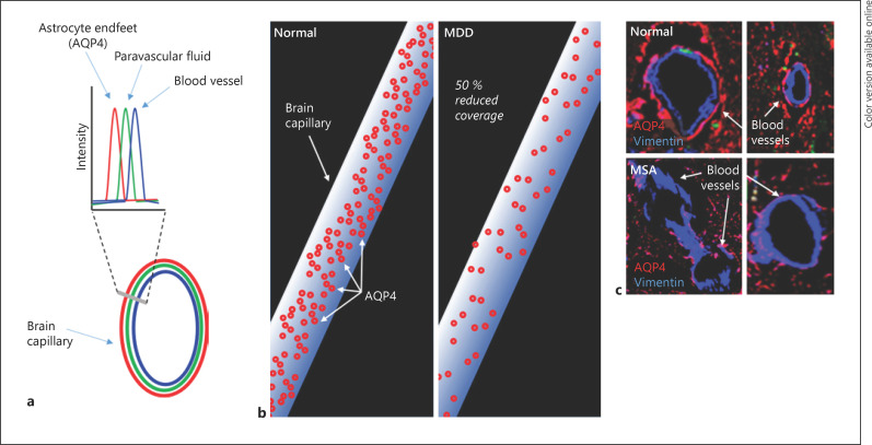
Fig. 1.
The glymphatic system and potential dysfunction in MDD and PD. a Glymphatic system structure shows solute (green) within the paravascular space [67] between the blood vessels (blue) and the AQP4-positive astrocytic endfeet (red). In normal brain, the glymphatic system likely clears solutes via the movement of CSF through these paravascular pathways. b The coverage of blood vessels by AQP4 associated with astrocytic endfeet in post-mortem brain showed a significant 50% reduction in the MDD group compared to the control group [69]. c Reduced AQP4 density around vimentin-positive blood vessels in the atypical PD, multiple system atrophy, compared to normal visual cortex (de Smet and Pountney, unpublished data). MDD, major depressive disorder; PD, Parkinson's disease; AQP4, aquaporin-4; CSF, cerebrospinal fluid; MSA, multiple system atrophy.

