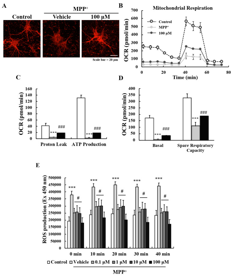
Figure 3.
EA reduced MPP+-induced oxidative stress and mitochondrial dysfunction in primary neurons. (A) Representative images showing that 100 µM EA pretreatment enhanced MMPs. Scale bar = 20 µm. (B–D) Oxygen consumption rates of primary neurons were measured using the Seahorse Cell Mito Stress assay to assess mitochondrial respiratory functions after co-treatment with MPP+- and EA+MPP+. Two independent experiments were performed, and results were averaged. Values shown are means ± SEs (n = 4). *** p < 0.001 vs. naïve controls and ### p < 0.001 vs. MPP+ controls (the analysis was performed using ANOVA with Fisher’s PLSD procedure). (E) Intracellular ROS levels were measured using DCF-DA dye at 10 min intervals. Values are means ± SEs (n = 8). *** p < 0.001 vs. naïve controls and # p < 0.05 vs. MPP+ controls (the analysis was performed using ANOVA with Fisher’s PLSD procedure).

