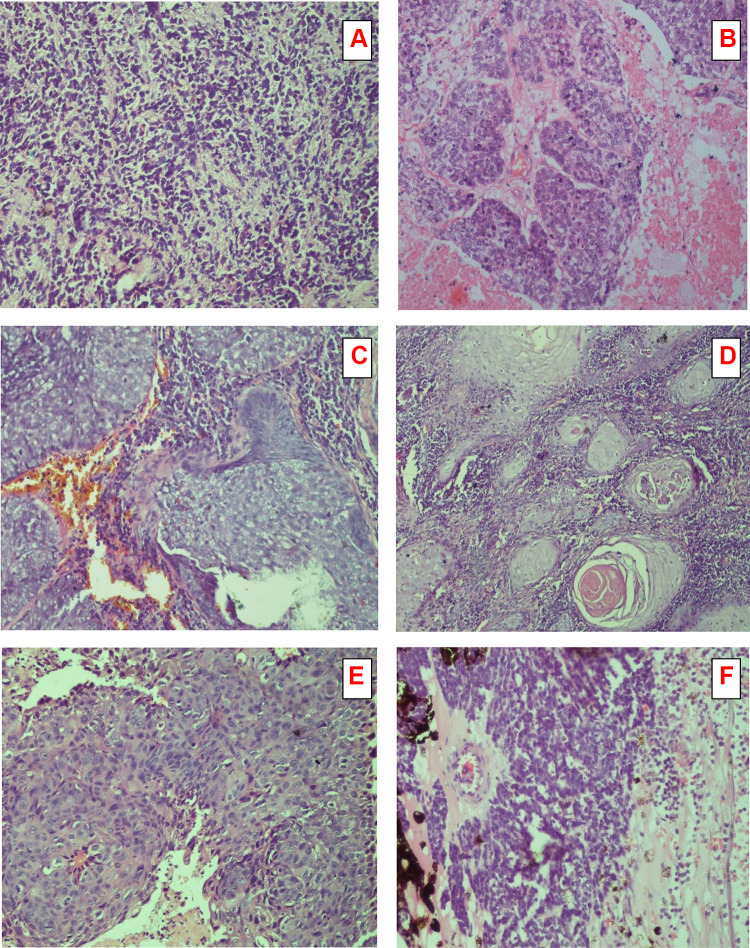
Figure 3.
Microphotograph of a different histological section stained (Hematoxylin and Eosin x 100 (X) showing (A). Rhabdomyosarcoma round cells (primitive rhabdomyoblast). (B). Sebaceous gland carcinoma moderately differentiated showing lobules of malignant cells with sebaceous differentiation. (C). Basal cell carcinoma showing nests of pigment laden atypical basal cells with peripheral palisading and mitotic figures. (D). Squamous cell carcinoma showing keratin pearls, numerous malignant cells, and mitotic figure. (E). OSSN showing keratin pearls and proliferation of irregular dysplastic epithelium with infiltration of sub epithelial tissue (F). Retinoblastoma well differentiated with Flexner-Wintersteiner rosette arrangement.

