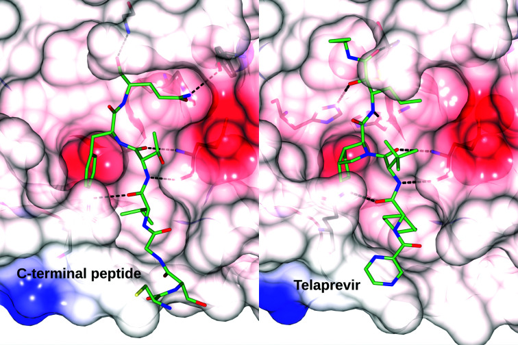
Figure 7.
Side-by-side comparison of the binding of the autoprocessed C-terminal peptide (PDB entry 7khp) and telaprevir (PDB entry 7k6d) in the active site of 3CLpro. Hydrogen bonds are shown as dashed lines. The protein molecule is represented as a semi-transparent charge-density surface with positive charge shown in blue, negative charge in red and hydrophobic character shown in white. The ligands in the active site are shown in stick representation with C atoms in light green, O atoms in red and N atoms in blue. 3CLpro residues forming hydrogen bonds to the ligands are shown as sticks under the charge-density surface.

