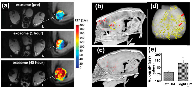
Figure 4.
(a) In vivo MR imaging of mice injected with iron oxide nanoparticle-loaded exosomes derived from melanoma cells. After 48 h footpad injection, the T1-weighted images and R2* mapping clearly suggested accumulation of exosomes in lymph node [42]. (b–e) In vivo CT imaging of Au nanoparticle-loaded exosomes after intranasal administration into mice with acute striatal stroke: (b) a CT image 24 h post-administration (the ischemic region is indicated by the yellow circle), (c) a CT image of control animal with saline injection, (d) ex vivo CT imaging and Au quantification within the animal brain 24 h post administration, and (e) CT analysis of Au density of left and right hemispheres (note: stroke was introduced in the right hemisphere) [32].*: (p < 0.05).

