. 2021 Feb 19;41(2):509–523. doi: 10.1148/rg.2021200049

Figure 11c.

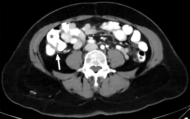
Optimization of window settings for virtual monochromatic (VMC) images. Axial portovenous phase abdominopelvic CT images obtained with iodinated oral contrast medium (*) show the bowel wall and folds (arrow). (a) A 120-kVp image at window width (WW) of 360 HU and window level (WL) of 60 HU. (b) A 40-keV VMC image at the same window settings shows significant "blooming" of contrast medium, hindering visualization of the bowel wall and folds. (c) A 40-keV VMC image at wider window settings (WW = 589 HU, WL = 88 HU) provides optimal display.

Optimization of window settings for virtual monochromatic (VMC) images. Axial portovenous phase abdominopelvic CT images obtained with iodinated oral contrast medium (*) show the bowel wall and folds (arrow). (a) A 120-kVp image at window width (WW) of 360 HU and window level (WL) of 60 HU. (b) A 40-keV VMC image at the same window settings shows significant "blooming" of contrast medium, hindering visualization of the bowel wall and folds. (c) A 40-keV VMC image at wider window settings (WW = 589 HU, WL = 88 HU) provides optimal display.