. 2021 Feb 19;41(2):509–523. doi: 10.1148/rg.2021200049

Figure 7c.

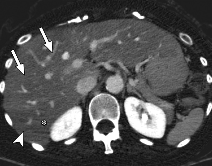
Beam hardening due to arm positioning. (a) Topographic image obtained for abdominopelvic CT with a dual-layer DECT scanner, which processes DECT images in the projection domain, shows that the patient was scanned in the "right arm down" position. (b, c) Axial conventional single-energy (b) and gray-scale MD-iodine (c) images show dark streaks (arrows) and bands (arrowhead) extending from the humerus, which degrade image quality and cause spurious lower attenuation and iodine distribution in the posterior hepatic segment (* in c).

Beam hardening due to arm positioning. (a) Topographic image obtained for abdominopelvic CT with a dual-layer DECT scanner, which processes DECT images in the projection domain, shows that the patient was scanned in the "right arm down" position. (b, c) Axial conventional single-energy (b) and gray-scale MD-iodine (c) images show dark streaks (arrows) and bands (arrowhead) extending from the humerus, which degrade image quality and cause spurious lower attenuation and iodine distribution in the posterior hepatic segment (* in c).