. 2021 Feb 19;41(2):509–523. doi: 10.1148/rg.2021200049

Figure 9a.

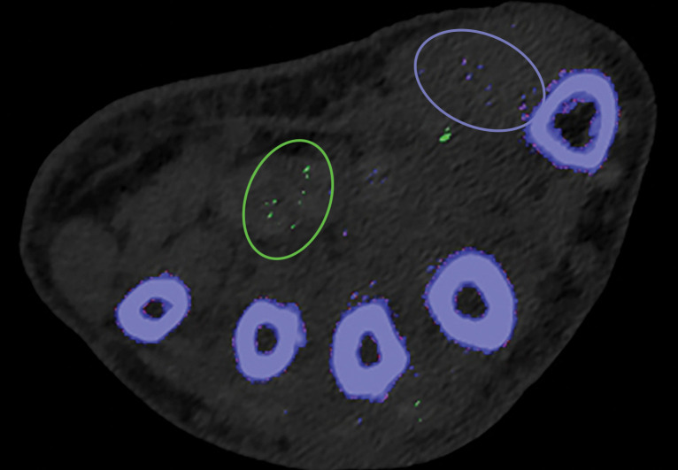
Appropriate kernel selection. MD-urate images of the wrist for gout evaluation with dual-source DECT. (a) Image processed with a bone kernel (Qr59d) shows artifactual green and blue pixels in the soft tissue that would indicate urate and calcium deposits, respectively (colors by default from the vendor). (b) On an image processed correctly with a soft-tissue kernel (Bf32d), these colored pixels have been removed and shown to be artifactual.

Appropriate kernel selection. MD-urate images of the wrist for gout evaluation with dual-source DECT. (a) Image processed with a bone kernel (Qr59d) shows artifactual green and blue pixels in the soft tissue that would indicate urate and calcium deposits, respectively (colors by default from the vendor). (b) On an image processed correctly with a soft-tissue kernel (Bf32d), these colored pixels have been removed and shown to be artifactual.